Navicular syndrome is one of the most frustrating of all hoof-related lameness problems, but modern management methods can help keep horses sound and pain-free, and improve their competition longevity.
“Navicular syndrome,
or caudal heel syndrome,
is a complex condition.”
Up until recently, navicular syndrome (NS) was referred to as navicular disease, but it is not a disease as such. Navicular syndrome, or caudal heel syndrome, is a complex condition that refers to the progressive deterioration involving both the navicular bone and arthritic changes associated with the coffin joint and pedal bone.
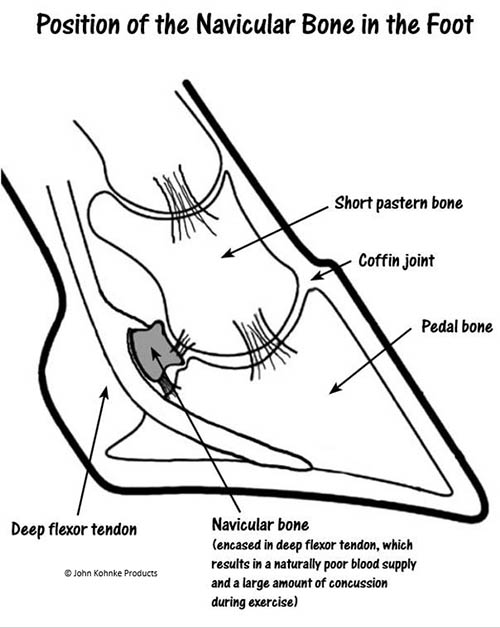
The navicular bone is a small bone, approximately 50mm long and as thick as the average small pinkie finger on a human hand. It acts as a pulley for the deep flexor tendon as it passes under the heel area to attach on the under surface of the pedal bone. When a horse accelerates, turns or stops suddenly on its heels, the navicular bone is loaded with extreme pressure, the blood supply is reduced, and it is concussed and compressed (or bruised) within the heel area.
RESEARCH INTO NS
Advanced diagnostic methods used by Dr William Widmer at Purdue University using magnetic resonance imaging (MRI) have provided new evidence that the underlying changes are associated with damage to soft tissue and cartilage, as well as bone demineralisation on the lower border of the navicular bone that articulates with the coffin joint. The overall result of navicular syndrome is lameness, which in time can become chronic.
Previously used X-ray diagnosis relied on classifying type, size and number of enlarged nutrient vessels or vascular channels, referred to as “lollypops”, on the lower border of the navicular bone. MRI scans have identified soft tissue from the joint (synovial) membrane inside the “lollypops” with the blood vessels being very small in horses with navicular syndrome. Sound horses have 3-5 cone-shaped synovial “holes” along the border of the navicular bone and up to 11% of “normal” horses have lollypop holes.
HORSES AT RISK
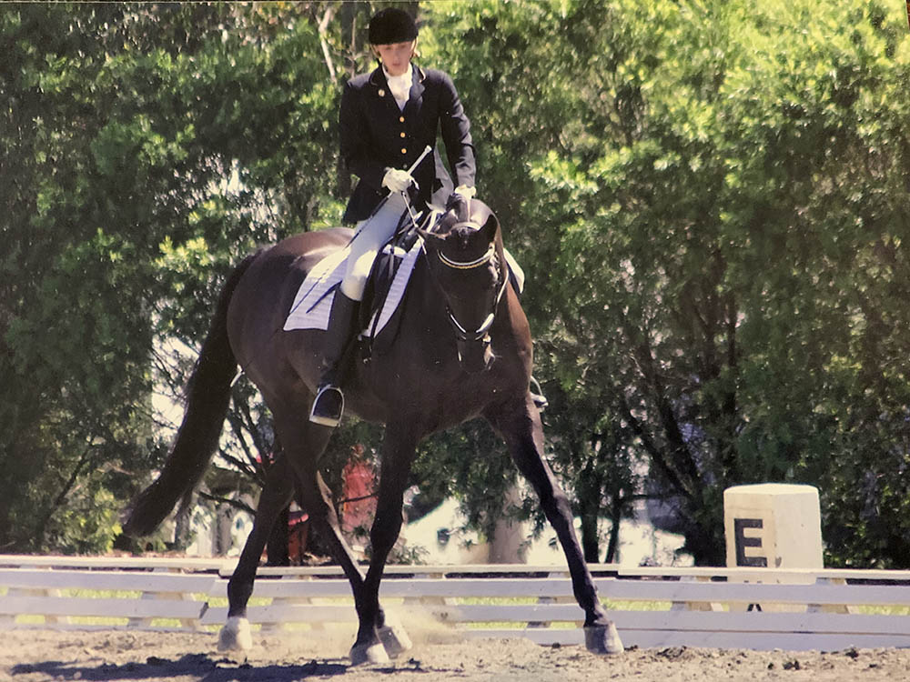
In most cases, one or both front hooves are affected — it is rarely seen in the hind hooves. Horses competing in highly physical equestrian disciplines aged between four and 15 years of age are most likely to suffer from NS due to the loading forces placed on the navicular bone and deep digital flexor tendon during heavy exercise and concussive loading. Whilst any breed of horse can develop the condition, research shows that certain breeds, such as Thoroughbreds, Quarter Horses and Warmbloods, are most at risk. Poor foot conformation in any horse, such as a long toe and low heels, also increases stress on the navicular bone and can contribute to the development of the condition.
COMMON SIGNS
NS exhibits early on as a subtle front limb lameness evident in one or both front limbs. It can be observed when a horse is led out of a stable or yard or during the warm-up portion of exercise. A horse’s natural, long, and free stride can become noticeably “choppier” or “scratchy” and less fluid in movement due to the horse stabbing the toe into the ground to take the pressure off the heel and back of the limb.
Usually, signs become less apparent after the horse is warmed up during the training session. It is also possible for signs of NS to be noticed in a cool-down session post work.
Early signs include an increased incidence of stumbling, developing a shuffling short stride or proppy gait in one or both front limbs, often transferring weight alternatively to the good limb. This short, straight-shouldered, more upright gait can be confused with a shoulder injury or a “tying up” in the shoulder during exercise. A horse’s reduction in willingness to perform may also be felt by the rider.
More advanced signs include wearing away the front toes more quickly as the horse attempts to land toe-first when exercising instead of the normal heel-first pattern at the walk or trot. An affected horse may drag the toes to minimise weight-bearing on the heel due to pain in this area. Affected horses will show a moderate level of lameness.
Chronic cases are characterised by the most obviously sign in a chronic form of lameness. Worn away and often chipped toes on the front hooves are also observed, as well as narrowing of the quarters and deepening of the frog sulci. This results in upright heel shape or a box-like appearance of the hoof on one or both limbs. Most horses have more developed signs in one front hoof.
“There has been
practical benefit from
supplementation.”
VETERINARY DIAGNOSIS
The diagnosis is a job for your vet. There are five main clinical methods used to identify progressive navicular bone changes, which include:
1. Assessment of front limb movement
An objective lameness exam on the circle lunge or under saddle is often the first step in identifying the change in front limb movement associated with caudal heel pain in a horse. Affected horses develop a shuffling, short-stepping gait, landing on the toe first rather than the normal heel-first ground contact when viewed from the side at a walk or trot. Many severely affected horses stand and shift weight from one limb to the other to ease the discomfort of weight-bearing on the heels.
2. Symmetrical examination of hoof shape
The typical chronic navicular-affected hoof has a boxy shape, with high narrow heels (as less weight is carried to expand the heels due to dull caudal heel pain), more upright quarters, sunken frog with deep bordering grooves (sulci) and short worn-away toes with evidence of toe “scrub” and wear due to landing toe-first and skidding on hard surfaces when exercising.
“MRI is now the
definitive method to investigate
the early changes.”
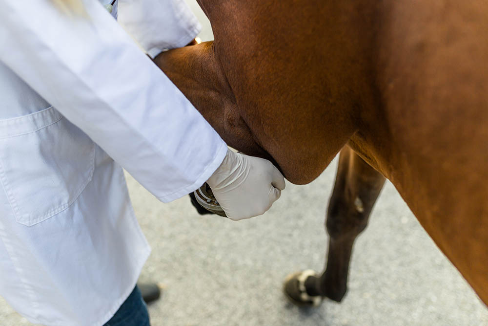
3. Pain on hoof tester application
It is important for a vet to assess how a possibly NS-affected horse perceives pain in the heel area. Many, but not all horses suffering from NS, will respond to the pressure of hoof testers.
4. Palmer digital nerve blocks
The palmer digital nerves (PDNs) in the mid-section on each side of the rear of the pastern provide the pain sensation to the heel area in a horse with caudal heel area pain. A horse with caudal heel pain will often trot off sound once the sensory nerves have been blocked.
5. Fetlock flexion tests
With the horse standing with one front hoof off the ground, the fetlock is bent back to the rear of the limb and held firmly for 30 seconds. On dropping the hoof to the ground, the horse is immediately trotted over 25-30 metres in a straight line. A horse with navicular pain will appear lame after the flexion test. If the caudal heel pain is relieved by the PDN block, the horse will trot off sound after four to five strides.
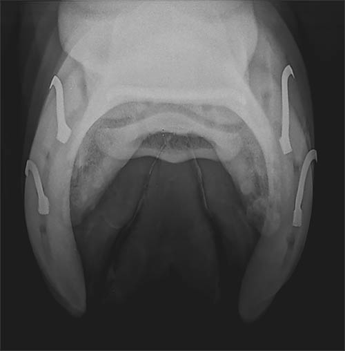
6. Navicular radiographs
Radiographs are taken from the front of the hoof wall in a downward slope to highlight the degree of navicular “lolly-popping” and bone changes within the navicular bone and the coffin joint.
7. Magnetic resonance imaging (MRI)
MRI is now the definitive method to investigate the early changes within the navicular bone. The early MRI studies found that navicular bones of horses with chronic hoof pain and lameness had increased fluid in the navicular marrow cavity, indicating an early inflammatory change. When the inflammation increases, it causes discomfort and load-bearing lameness, and it can eventually burst out of the back of the navicular bone to adhere to the deep flexor tendon, resulting in bony changes within the joint structure of the coffin joint and resulting in severe chronic lameness.
“Kohnke’s Own Redi-Flex
is a comprehensive
joint supplement.”
MANAGEMENT OF NS
Unfortunately, navicular syndrome is a chronic and degenerative condition, therefore it cannot be cured. However, modern management methods are effective in helping to keep horses sound, pain-free, and can increase competition longevity. In recent years, the prognosis has improved significantly due to the increased use of MRIs as well as the communication between farriers and veterinarians. The severity of clinical signs, the horse’s age, and intended use (including workload), are important considerations when developing a management plan. Younger horses with less severe clinical signs are the most suitable candidates for traditional therapy.
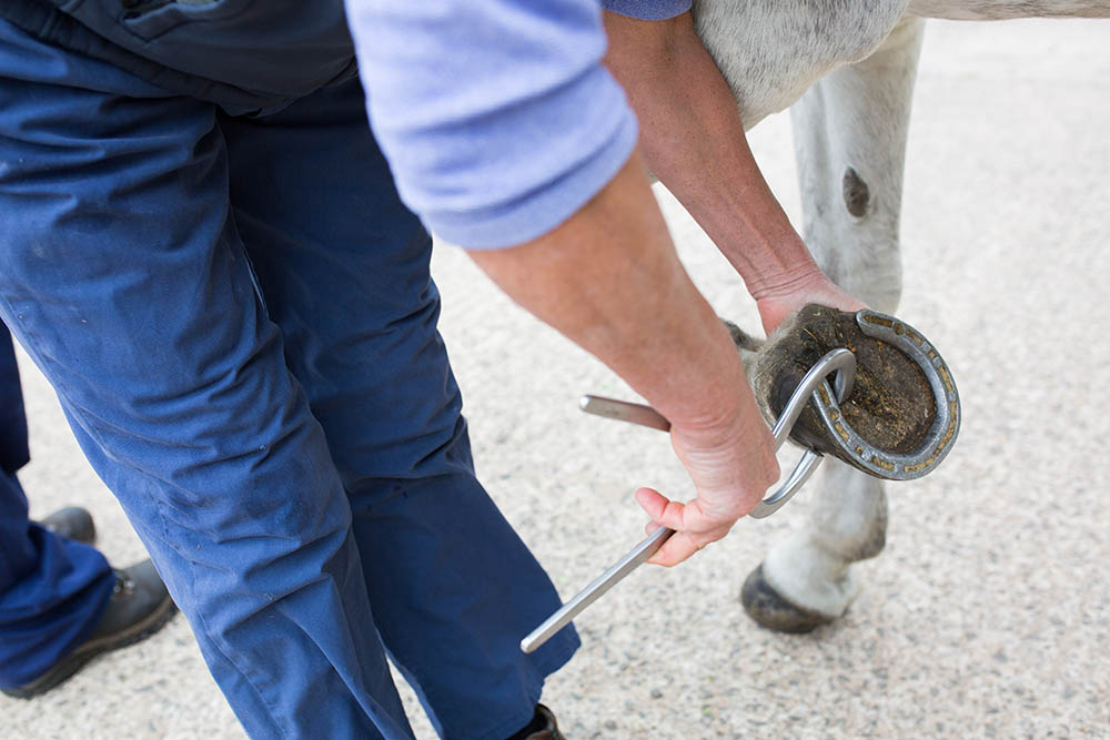
Non-surgical treatments include rest, corrective and therapeutic trimming or shoeing, as well as medical therapies including use of anti-inflammatories and other veterinary prescribed medications.
The biggest focal point in non-surgical treatment of NS is corrective shoeing. Careful evaluation of foot conformation and balance is key. Forces placed on the navicular region, including strain on the deep digital flexor tendon, can be reduced to help ease heel pain and lessen the severity of the condition. X-ray images of hoof angles are especially beneficial for farriers to assist with corrective shoeing.
As NS is a progressive degenerative problem within the navicular bone and its fibro-cartilage border, pain relief through the use of anti-inflammatory drugs should be discussed with your veterinarian. Long-term use of anti-inflammatory drugs is illegal in many equestrian competitions and can also have adverse health effects, especially on the digestive system.
The deposition of long-acting anti-inflammatory synthetic corticosteroid injections, into the two sacs of the navicular bursa within the coffin joint, is helpful to provide temporary relief from the constant pain and lameness of NS. A horse with a positive fetlock flexion test may benefit in the short term from a corticosteroid injection into the navicular bursa to reduce the inflammation. This can provide relief within 5-7 days and, once the horse is sound, it is considered best to put it back into light work to restore hoof circulation and weight-bearing.
There has also been the development of other new therapies for treatment. The use of injectable bisphosphonates, as recommended by veterinarians, has been useful to help improve bone density, increasing the capacity of the navicular bone to withstand mechanical loading in an exercising horse. This may reduce the risk of navicular fracture due to bone decalcification or osteoporosis, for example, in an aged jumping horse as it lands heavily on its front hooves on to a compacted surface.
The use of high-intensity sound waves or extracorporeal shock wave therapy (ECSWT) has been trialled to help relieve pain and shorten healing time of collagen tissue in the ligament attachments to the navicular bone within the coffin joint. It can especially be useful in conjunction with bisphosphonate therapy for bone repair.
“Good management
methods are key to reducing
the risk of NS.”
BENEFITS OF A DAILY JOINT SUPPLEMENT
There has been practical benefit from supplementation with an equine joint supplement to help restore the environment and health of the articular surfaces of the coffin joint.
There are myriad joint supplements available, as they are the most common supplement given orally to exercising and aged horses. Kohnke’s Own Redi-Flex is a comprehensive joint supplement with over 10 high-potency, joint-active nutrients that benefit joints, ligaments and tendons. Trials during the development of Redi-Flex included case studies of horses that had been diagnosed with NS. Horses showed improvement in their gait and stride length with increased willingness of movement and reduced lameness.
Redi-Flex can be considered a complementary supplement to other management used to help NS horses return to work or lead a relatively pain-free and active life as they age.
SUMMARY
Although navicular syndrome cannot be fully cured, proper management and treatment can help to reduce stress, inflammation and pain in the affected areas, helping to prolong the competitive longevity of the horse.
Good management methods are key to reducing the risk of NS. These include correct and regular hoof care, proper nutrition and weight management, a specifically tailored, regular exercise schedule for each horse, as well as supportive footing on exercise surfaces. EQ
(Dr John Kohnke BVSc, Roseworthy Diploma of Agriculture, is the founder of Kohnke’s Own, while Georgia Grech BSc (Zoology) is one of the company’s nutritional advisors, marketing and social media manager and a competitive eventer.)
“My beloved dressage horse Freddie was 14 years old when he was diagnosed with navicular syndrome. It was a heartbreaking moment as I realised what this meant for Freddie’s future as a dressage horse. We initially tried Pentosan, which helped along with corrective shoeing and a good management plan that kept him competing for a few more years. Freddie was retired when he was 18, as I felt he just didn’t have great freedom of movement through his shoulder anymore and the vet had advised that there was nothing else that could be done. When the trials for Redi-Flex began, Freddie was no longer on a joint supplement and we were struggling to maintain paddock soundness. He was very stiff and even lame on cold mornings through winter. He was started on the loading dose of the Redi-Flex trial and within two weeks I noticed an improvement. It has been almost four years since Freddie was started on Redi-Flex and 10 years since he was diagnosed with navicular syndrome. Freddie is currently walking, trotting and cantering around his paddock happily and I am so glad that I still have this special horse in my life.” — Sian R