A hernia is the protrusion of an organ, or part of an organ, through an opening in the wall of the cavity that normally encases that organ. There are several types of hernias that can affect horses — some are relatively common, such as umbilical hernias seen in foals, and others are rare, such as diaphragmatic hernias.
“(Umbilical hernias) are relatively
common abnormalities in foals.”
Most types of hernia usually involve the organs that are situated in the abdomen and they are categorised by the area of the cavity that the organs protrude through. For instance, an inguinal hernia is a hernia associated with abdominal contents coming through the inguinal ring.
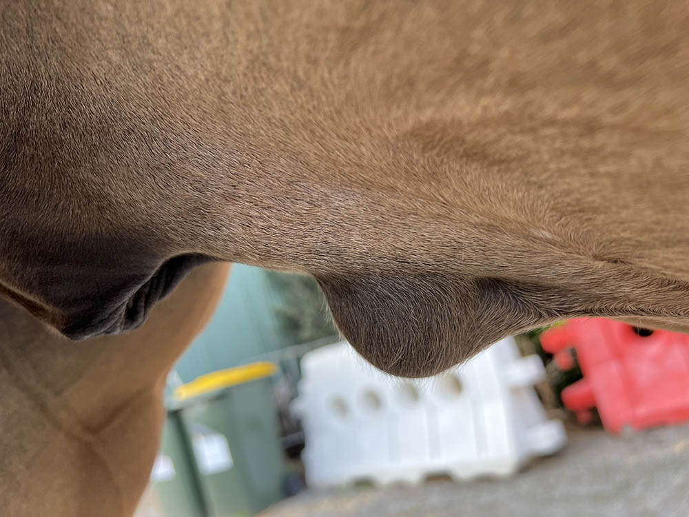
Umbilical hernias are “true” hernias in that the herniation occurs through a normal or congenitally enlarged opening and the contents within the hernia are contained within a hernial sac. They are relatively common abnormalities in foals that occur around the time of birth, although they are not always noticed by the owner initially, as the contents within the hernia may be minimal at birth but become more visible during the first few weeks of life. They can fluctuate in size, depending on the abdominal content that moves in and out through the hernial ring.
Umbilical hernias vary in size and are commonly classified by vets according to the number of fingers that can be placed up through the defect in the abdominal wall. In my experience, most hernias are around the 2 to 3 finger size, relating to about 4-6cm, however, occasionally the abdominal defects are much larger, and sometimes much smaller, allowing just a single finger into the hole.
Whilst some hernias will contain part of the intestinal tract, other times there will be omentum (a fatty layer of tissue that is part of the peritoneum), fat and abdominal fluid present. These hernias infrequently cause the horse discomfort, however, there is a risk in leaving them untreated as a piece of intestine can become caught in the sac causing severe colic. Once entrapped, surgery is required to free the intestine, and in more serious cases to remove the compromised piece of bowel that has been strangulated by the hernial ring.
There are a few treatments that have been used to remove umbilical hernias and they each have their advantages and their disadvantages. The gold standard for treatment is to replace the herniated tissue and surgically repair the gut wall defect under general anaesthesia. The disadvantage of doing surgery is that, if for any reason the surgical wound breaks down, the abdomen is open and there is an increased risk of peritonitis occurring.
Another form of treatment that has been used to “fix” umbilical hernias is the placement of an elastrator ring on the hernial sac. This does not repair the defect in the gut wall but removes the space that the abdominal contents can move into. This has been done using general anaesthesia and performed as a standing procedure. I do not agree with the practice of applying a ring to a hernia in a standing horse as there is a significant risk of a portion of the hernia contents becoming trapped in the elastrator ring and this can be fatal.
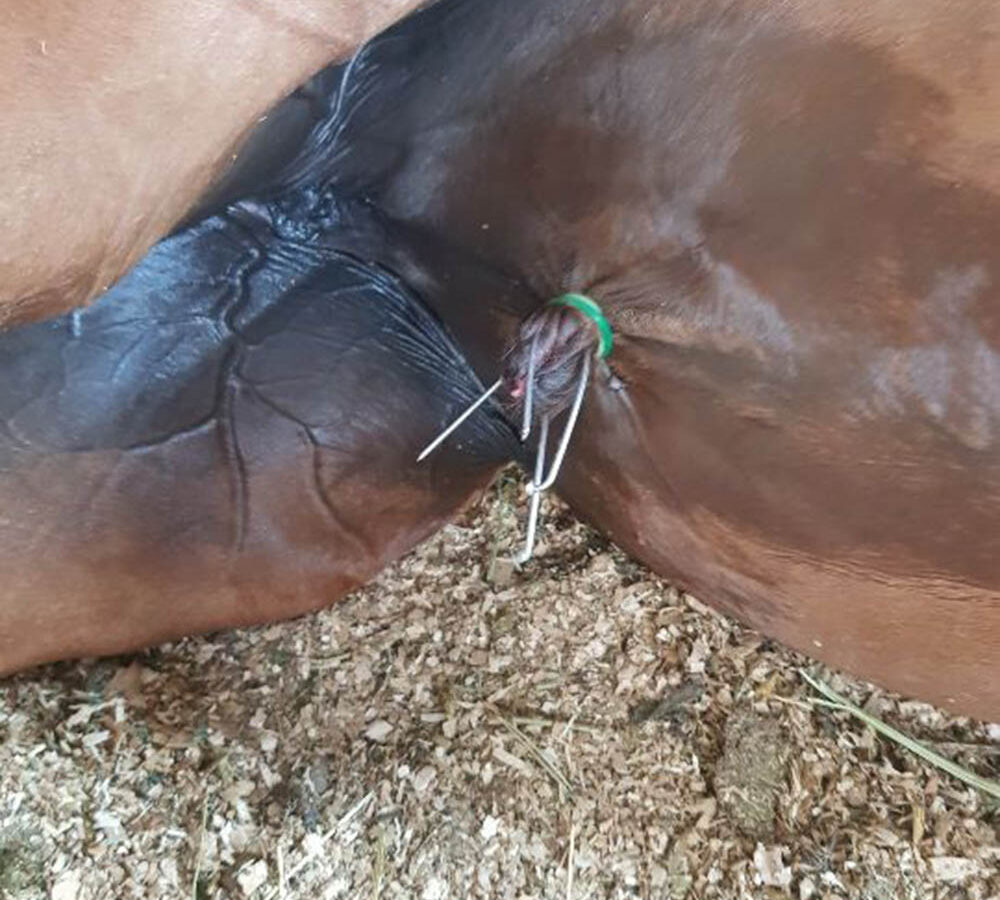
The risk is reduced when the horse is anaesthetised and positioned on its back, as gravity usually ensures the abdominal contents fall back down into place, allowing the ring to be placed over the skin and hernial sac. The hernial sac and skin that are contained within the elastrator ring slowly die due to lack a lack of blood supply and drop off after 3 to 4 weeks, leaving a small scar on the lower abdomen.
INGUINAL HERNIA: These are predominantly congenital defects and are not common, but if they occur, are seen in newborn male foals. Inguinal hernias are thought to be inherited conditions, with Standardbreds one of the more susceptible breeds. These hernias can involve either one or both inguinal rings. Herniation occurs due to an enlarged ring or opening between the gut and the scrotum that allows the intestines to move freely out of the abdomen into the scrotal region around the time of birth. When the intestines move into the scrotum, they are commonly referred to as a scrotal hernia.
For those inguinal hernias where the intestines remain within the vaginal process or extension of the peritoneal cavity (the lining of the abdomen), conservative management and time can often result in a resolution without the need for surgical intervention. The hernia needs to be manually reduced daily to encourage the intestines to move back into the abdomen and hope that the vaginal ring decreases in size relative to the abdominal contents, thus reducing the risk of the hernia returning. In hernias that occur when the vaginal process tears, usually during foaling due to increased abdominal pressures, the intestines move into the subcutaneous tissues, causing a life-threatening situation that requires urgent veterinary attention.
If surgery is not performed there is a huge risk that the intestines will overstretch the fragile scrotal skin and cause it to ulcerate and tear, exposing the intestines to the environment. Once the intestines are exposed, they are easily damaged or infected, ultimately causing death. During surgical repair of these scrotal hernias, the testicle on the side of the hernia is usually removed, and the open ring sutured closed to eliminate the risk of more intestine moving through it.
When the hernias are left to resolve on their own, castration should be performed when the foal is older as the condition is heritable, and these colts should not be used for breeding purposes. It should be noted, however, that castration carries a risk of evisceration or loss of intestines through the castration site, and it is strongly recommended that any foal with a history of congenital inguinal herniation undergoes a closed castration procedure to minimise the risk of the evisceration. A closed castration is one where the scrotal vessels and tunic is ligated and closed to prevent any abdominal contents moving out through the opening.
Adult male horses can have what is termed an “acquired inguinal hernia”, where some form of strenuous physical activity causes the gut contents to move out through the inguinal ring and into the scrotum. There is some thought that the vaginal ring in these horses is congenitally enlarged and therefore the stallion is predisposed to herniation. Clinically, the stallion will show signs of colic and careful observation will identify a swollen and painful scrotum. There are reports of these hernias being fixed conservatively, however, if there is any evidence that the intestines within the scrotum have been seriously compromised, immediate surgery is required to assess the intestines and repair any damage.
There have been infrequent cases of mares or geldings acquiring an inguinal hernia, however, these are rare as the inguinal ring in mares and geldings are smaller and don’t allow the intestines to move through them.
“False” hernias differ from the true hernias mentioned above in that they occur because of a tear or abnormal hole in a surface that allows an organ or part of an organ to move out through the opening but there is no hernial ring and the contents are not contained within a hernial sac. They are less common than the true hernias and can result in many different clinical signs, depending on what has herniated and where the hernia is. Below are listed some of the various false hernias that can be seen in horses.
INCISIONAL HERNIAS: These occur when a surgical wound into a cavity breaks down and the contents of the cavity bulge out under the skin or subcutaneous tissues, causing a visible soft swelling. The more common incisional hernias occur following an abdominal laparotomy, where circumstances have resulted in the internal sutures weakening and the abdominal contents protruding through the gut wall and sitting on the ventral abdomen.
Multiple abdominal surgeries can predispose a horse to an incisional hernia as the wall is weakened when it is opened and sutured more than once, as can occur with some colic patients that require the surgeons to re-operate when complications occur following the initial surgery. Infection can also lead to wound breakdown and an ensuing hernia. Sometimes a surgeon will apply a body bandage or wrap to the abdomen to try and minimise the risk of the wound breaking down and a hernia occurring.
TRAUMATIC INDUCED HERNIAS: These can occur through the gut wall when the internal muscles are weakened or damaged physically, allowing the abdominal contents to spill out into the subcutaneous tissues, forming a hernia. They are visible as soft fluctuant swellings on the abdominal wall and, depending on their size and location, can be treated either conservatively or surgically.
DIAPHRAGMATIC HERNIAS: Diaphragmatic hernias are rare in horses and can be congenital or acquired. The defect in the diaphragm allows the intestines to go through into the pleural cavity and can result in colic symptoms or respiratory symptoms, depending on how large the defect is and what herniates through. Sometimes only a small hole exists and the intestine that passes through is strangulated, causing severe colic with minimal effect on the respiratory system. In other instances, the hole is large, allowing the intestines to become displaced into the pleural cavity, greatly decreasing the room available for the lungs to expand, resulting in the horse having difficulty breathing. They are usually diagnosed with the ultrasound machine as abdominal contents are visualised in the chest cavity, however, in some cases the abdominal contents are not visible, and surgery is required to diagnose the problem. The success of the surgery will depend on the size and location of the defect in the diaphragm as not all areas of the diaphragm are accessible to the surgeon and euthanasia may be required.
HIATUS HERNIA: A very rare hernia that occurs when the stomach, or part of the stomach, protrudes up through the diaphragm and causes clinical symptoms related to eating and recurring choke episodes. These can be managed conservatively but management changes when the horse eats.
PERINEAL HERNIA: Also very rare, with a single case being reported in a donkey, although more common in other breeds of animals. The perineum in the horse is the area around the anus and vulvar lips (mares) and occurs when the pelvic musculature is damaged or weakened, allowing organs in the hind part of the abdomen to herniate. They appear as soft swellings on one side or the other of the perineum and the hernia can contain intestines, bladder, or rectum. In the case of this donkey, surgery was performed, and the donkey made an uneventful recovery. EQ
YOU MIGHT ALSO LIKE TO READ THE FOLLOWING BY DR MAXINE BRAIN:
Osteochondromas: Benign But Irritating – Equestrian Life, February, 2022
Don’t Forget the Water – Equestrian Life, January, 2022
Understanding Anaesthesia – Equestrian Life, December, 2021
A Quick Guide to Castration – Equestrian Life, November, 2021
Caring for Mammary Glands – Equestrian Life, October, 2021
Sepsis In Foals – Equestrian Life, September 2021
Understanding Tendon Sheath Inflammation – Equestrian Life, August 2021
The Mystery of Equine Shivers – Equestrian Life, July 2021
Heads up for the Big Chill – Equestrian Life, June 2021
The Ridden Horse Pain Ethogram – Equestrian Life, May 2021
The Benefits of Genetic Testing – Equestrian Life, April 2021
Heavy Metal Toxicities – Equestrian Life, March 2021
Euthanasia, the Toughest Decision – Equestrian Life, February 2021
How to Beat Heat Stress – Equestrian Life, January 2021
Medicinal Cannabis for Horses – Equestrian Life, December 2020
Foal Diarrhoea Part 2: Infectious Diarrhoea – Equestrian Life, November 2020
Foal Diarrhoea (Don’t Panic!) – Equestrian Life, October 2020
Urticaria Calls For Detective Work – Equestrian Life, September 2020
Winter’s Scourge, The Foot Abscess – Equestrian Life, August 2020
Core Strengthening & Balance Exercises – Equestrian Life, July 2020
The Principles of Rehabilitation – Equestrian Life, June 2020
When is Old, Too Old? – Equestrian Life, May 2020