The umbilical cord is the anatomical structure responsible for maintaining the life of the foal in utero by connecting it to the mother via the placental attachment to the uterus. Upon birth, its juncture with the foal usually seals naturally – but not always.
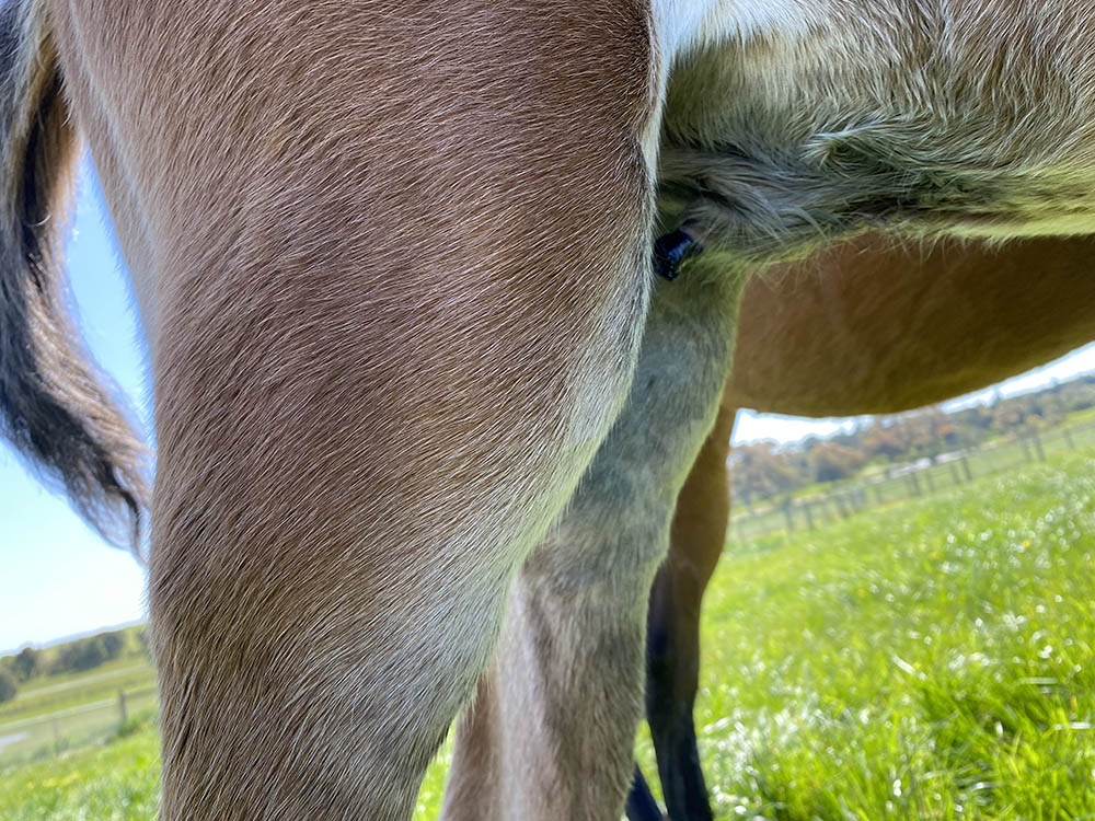
“The owners are alerted to a problem
when they see a wet area on the abdomen.”
The umbilical cord contains two arteries, and a vein, that are responsible for transferring nutrients and oxygen and deoxygenated blood via the bloodstream between the mare to the foal. The cord also contains the urachus, a thin-walled tube connecting the foetal bladder to the sac around the foal, known as the allantois. The urachus allows foetal waste, in the form of urine, to be expelled from the foetus into the fluid of the allantois, which is the outer of the two sacks of fluid that the foal is suspended in.
During normal parturition, the umbilical cord spontaneously ruptures shortly after delivery, sealing this opening between the foal and the outside environment. Once the umbilicus closes, it quickly becomes shrivelled, remaining as a dried-out structure on the lower abdomen for several weeks before dropping off. When this process doesn’t occur correctly, or there are complicating factors that interfere with it, the foal is at risk of developing serious infections, some that can be severely debilitating and some that can be fatal.
Complications include the incomplete closure of the urachus, allowing urine to continue to leak from the cord; excessive bleeding from one or more of the blood vessels in the cord; and systemic infections and local trauma that compromise the health of the umbilicus. Frequently, the result of any impediment to the normal rupture and subsequent closure of the umbilical cord is infection.
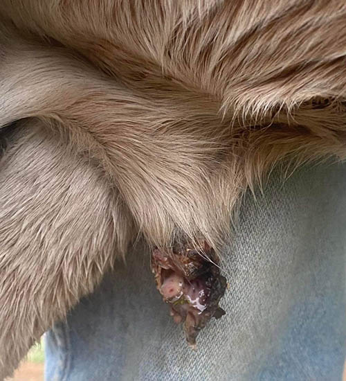
A “patent urachus” is the medical term given to the incomplete closure of the urachus, allowing urine to escape from the bladder and leak out through the umbilical stump. The urachus connects directly to the apex of the bladder, and failure of this structure to close and seal results in either the continuous dripping/dribbling of urine from the umbilical stump, or the foal posturing and passing a stream of urine from the umbilicus. The owners are alerted to a problem when they see a wet area on the abdomen, or they see fluid dripping or being urinated from the umbilicus.
PRIMARY CONDITION
Patent urachus can occur as a primary condition (sometimes referred to as a persistent urachus), or it can be acquired in the days to weeks after foaling following excessive straining, prolonged periods of recumbency, or occasionally following increased abdominal pressure applied in the vicinity of the bladder. Excessive straining can occur in foals with retained meconium or those that have been administered multiple enemas, causing an irritation to the rectum that induces straining. Prolonged lying on the ground is thought to increase the internal abdominal pressures, forcing urine out through the urachus, but it could be seen more frequently in foals that are recumbent for long periods, as these are often the foals that have systemic infections or compromised immune systems and so are predisposed. Increasing intra-abdominal pressures can occur when foals are lifted from the ground by people holding them tightly around the abdomen or by using slings.
A primary patent urachus that is present from birth is thought to be more prominent in foals with an unusually long umbilical cord, as these are prone to torsion in utero, causing increased pressures and dilations in the urachus. Dilations are enlarged sections of the urachus that occur when the fluid (urine) within the tube cannot flow out and cause the walls to expand under pressure
Regardless of whether the patent urachus is primary or secondary, foals require broad-spectrum antibiotics to prevent infection, or if already infected, to treat infection that is present. Veterinarians have tried applying chemicals to cauterise the cord in order to close the opening. This is the application of strong antiseptics or chemicals designed to cause swelling and inflammation of the cord in order to seal it, however, this can backfire and cause too much destruction to the cord, leaving the cord damaged and more susceptible to infection.
Instead, the preferred method is to use more gentle antiseptic solutions such as chlorhexidine to clean the area without causing more destruction. Povidine Iodine sprays (0.1%) are safe to use but please note, these are much more diluted than the strong iodine solutions that are available and which will cause cell destruction.
In a small percentage of cases with a patent urachus, closure of the urachus does not occur with conservative treatment alone and surgical intervention is required. This involves opening the abdomen, ligating and removing the persistent piece of urachus to resolve the problem.
INFECTED UMBILICAL CORD
“Omphalitophlebitis” is the term used for an infected umbilical cord and this can occur at or around the time of birth or develop in the days to weeks following birth. This involves an infection that also involves the blood vessels and not just the urachus. The umbilicus can become enlarged and swollen and this is a key indication that something is wrong, however, the absence of swelling and a discharge externally does not mean that the umbilicus is not infected.
It is possible to have an infection occurring with the abdominal remnants of the cord that is not immediately apparent to the owner, and any indication of an unexplained fever, lethargy or off suck scenario warrants a closer examination of the umbilicus. It is important to seek veterinary treatment if the umbilical stump becomes swollen, as untreated infections can seed infection to other areas of the body; often to joints causing joint ill, a life-threatening infection that can result in severe lameness and, sadly, death.
Foals with omphalitophlebitis are usually treated with broad-spectrum antibiotics until the cord returns to normal, but occasionally these infections can persist or develop into abscesses, requiring more intense treatment and surgical intervention.
In a small number of foalings, the rupture of the umbilical cord does not occur correctly and tears off unpredictably, causing the umbilicus to bleed profusely. If the bleeding doesn’t stop quickly, the foal can lose a substantial amount of blood. In some circumstances, foals require medical intervention to stop the bleeding, and this is usually the application of an umbilical clip or a ligature around the cord to stop the bleeding. Whilst this is sufficient to prevent further blood loss, a clip or ligature can also increase the risk of an infection occurring, so the umbilical stump should be monitored for signs of swelling or infection, so any infection can be treated quickly.
In most cases, the clip or ligature will fall off when the dried umbilicus falls off, but clips can be removed earlier to reduce the chances of them being caught and prematurely ripped off on objects in the environment. On a rare occasion, a foal may require fluid therapy or a blood transfusion if bleeding has been continuous for a long period, but in my experience, this would be very rare.
Tears in the urachus within the abdomen of the foal can allow urine to leak directly into the abdomen, rendering the foal quite ill. Foals become lethargic and can show abdominal distention, electrolyte disturbances and neurological signs if not detected early. If urine is leaking into the abdomen, surgery is recommended as soon as the foal is stabilised and able to have anaesthesia.

ULTRASOUND EXAMINATION
Regardless of the type of concern or issue with the umbilicus, an ultrasound examination of the umbilical area is highly recommended in all but the simplest cases. Using the ultrasound allows the vet to follow the blood vessel forward towards the liver to look for thickenings, infections and tears. The urachus can be tracked back towards the bladder to look for dilations in the urachus that may not be evident otherwise, or tears in the structure that can result in urine escaping into the abdomen via the urachus if it fails to seal properly. The detection of internal abscesses that have developed without any external indications is extremely valuable, as these abscesses can get very large and if not diagnosed and treated promptly, lead to major health concerns.
It is vitally important that any foal showing abnormalities pertaining to the umbilicus in the first few weeks of life has a blood test to measure its IgG levels. This is to ensure that the foal has ingested sufficient colostrum or received adequate immunoglobulins parenterally to give it some protection from infection. Failure of passive immunity (failure of the foal to receive adequate colostrum) can be a problem in the healthiest of foals, but when it occurs in conjunction with an umbilical infection, the likelihood of septicaemia, pneumonia and joint infections increases greatly. Any foal with a compromised umbilicus that has insufficient levels of immunoglobulins when tested, should be treated with intravenous plasma to increase its chances of overcoming an infection. The vet may also perform other blood tests to help them rule in or out problems such as septicaemia.
Umbilical hernias are swellings noted on the lower abdomen at the site of the umbilicus, but are caused by the incomplete closure of the abdominal wall where the umbilicus was attached and are not for the purpose of this article discussed as an umbilical cord problem. EQ
YOU MIGHT ALSO LIKE TO READ BY DR MAXINE BRAIN:
Retained Foetal Membranes – Equestrian Life, October 2022
Preparing for Laminitis – Equestrian Life, September 2022
Working Together for Best Outcomes – Equestrian Life, August 2022
What Constitutes an Emergency – Equestrian Life, July 2022
Peri-Tarsal Cellulitis Calls for Quick Action – Equestrian Life, June 2022
Sinusitis: Not To Be Sneezed At – Equestrian Life, May 2022
Japanese Encephalitis: No Cause For Alarm – Equestrian Life, April 2022
Hernia Learning Curve – Equestrian Life, March 2022
Osteochondromas: Benign But Irritating – Equestrian Life, February 2022
Don’t Forget the Water – Equestrian Life, January 2022
Understanding Anaesthesia – Equestrian Life, December 2021
A Quick Guide to Castration – Equestrian Life, November 2021
Caring for Mammary Glands – Equestrian Life, October 2021
Sepsis In Foals – Equestrian Life, September 2021
Understanding Tendon Sheath Inflammation – Equestrian Life, August 2021
The Mystery of Equine Shivers – Equestrian Life, July 2021
Heads up for the Big Chill – Equestrian Life, June 2021
The Ridden Horse Pain Ethogram – Equestrian Life, May 2021
The Benefits of Genetic Testing – Equestrian Life, April 2021
Heavy Metal Toxicities – Equestrian Life, March 2021
Euthanasia, the Toughest Decision – Equestrian Life, February 2021
How to Beat Heat Stress – Equestrian Life, January 2021
Medicinal Cannabis for Horses – Equestrian Life, December 2020
Foal Diarrhoea Part 2: Infectious Diarrhoea – Equestrian Life, November 2020
Foal Diarrhoea (Don’t Panic!) – Equestrian Life, October 2020
Urticaria Calls For Detective Work – Equestrian Life, September 2020
Winter’s Scourge, The Foot Abscess – Equestrian Life, August 2020
Core Strengthening & Balance Exercises – Equestrian Life, July 2020
The Principles of Rehabilitation – Equestrian Life, June 2020
When is Old, Too Old? – Equestrian Life, May 2020