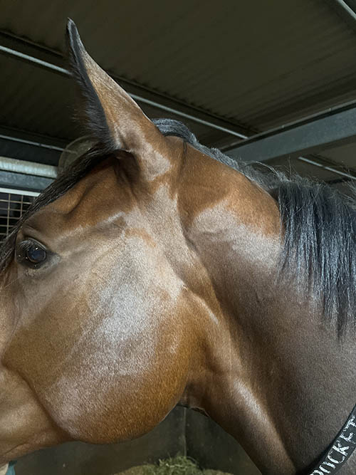
Salivary glands are not often a topic of discussion, medically speaking, as they do not cause the horse many problems when compared to other structures in the mouth or the gastrointestinal tract. They are, however, an integral part of the gastrointestinal tract.

A horse has three pairs of salivary glands, the parotid, the mandibular and the sublingual glands. The parotid glands are the largest and the most visible of the three. They sit either side of the head, just below the ear, extending from the mandible to the first neck vertebra (atlas) and are around 20-25cm in length in the average 500kg horse.
The parotid gland is closely associated with many important structures and vessels in this area including the guttural pouch, the jugular vein, various nerves and part of the external carotid artery. A duct carries saliva from the gland down into the mouth, where it is then released via a small papilla near the upper third cheek tooth.
The mandibular salivary gland is smaller than the parotid and is concealed from view by the overlying parotid gland and the lower jaw. The mandibular duct similarly transports saliva from the gland into the mouth, where it is released closer to the canine tooth.
The sublingual salivary gland is located within the mouth between the tongue and the inside of the mandible. This gland differs from the parotid and the mandibular glands in that it doesn’t connect to a single duct to transport the saliva but rather has a series of small ducts (about 30) that open onto a small papilla to release saliva.
The release of saliva into the oral cavity is stimulated by the chewing motion, with increased amounts released as the horse chews longer and more frequently. This is seen commonly when hay is fed as opposed to grain feeds and is thought to be one of the reasons why horses prefer to eat hay over grain when they suffer with ulcers.
Unlike some mammalian species such as humans and dogs, there is little to no enzyme activity in the saliva to help digest the feed and its role in the digestive process is mechanical and not metabolic. Saliva serves to soften and lubricate the food to allow it to pass easily down the oesophagus into the stomach. It also contains high levels of sodium bicarbonate that acts as a buffer to increase the pH level in the stomach by neutralising some of the acids produced in the stomach that we know can lead to gastric ulcers. Other components of saliva include calcium, potassium, chloride, glycoproteins, and polysaccharides.


There are a couple of medical conditions that can affect the salivary glands, but overall, these are uncommon. Swelling of the parotid gland should be differentiated from swelling of underlying tissues such as the guttural pouch or lymph nodes in the throat area, as these structures, if enlarged, can make the parotid gland bulge out, even though it is clinically unaffected.
Sialoliths are stones that form in the salivary gland or duct and can cause a blockage as they move through the duct. They are more commonly seen in the parotid duct but have been found on the odd occasion in the mandibular duct and rarely if ever associated with in the sublingual salivary glands, as these salivary glands do not have a common duct as stated above. The distal or lower part of the parotid duct is the most common site for sialoliths to be located, however, they can be formed in the proximal parotid duct, where they are more difficult to detect. In the distal part of the parotid duct, these stones can sometimes be palpated as small, hard moveable lumps on the outside of the cheek. They are typically composed of calcium deposits that start off surrounding a small particle or nidus and continue to enlarge with further deposits of calcium products added in layers over time.
Sialoliths can block the flow of saliva through the duct, causing it (saliva) to bank up and distend the duct from immediately behind the stone back towards the gland. These soft, fluctuant swellings can be quite distinct when they occur along the portion of the duct that passes under the jaw and across the masseter muscle (the large muscle on the side of the jaw) and take on a sausage-like appearance where the duct is dilated. These dilated ducts can rupture, spilling saliva into the subcutaneous tissues, forming an accumulation of saliva or mucocele in the subcutaneous tissues.
Sometimes the duct can rupture and drain out through the skin causing a persistent dripping of saliva until the wound eventually seals over. Other clinical signs that may be seen with sialoliths include ulceration inside the cheeks due to rubbing against the stone, quidding or dropping semi-chewed food due to reduced lubrication, difficulty swallowing due to lack of lubrication and facial paralysis if the sialolith is pressing on the facial nerve.
Sialoliths can be diagnosed either by direct palpation, radiographs or ultrasonography, depending on their location along the duct, with ultrasonography the better option if available. For complicated cases, a CT of the skull can reveal the site and size of the stones.
Removal of the sialoliths surgically, either through the mouth or through the outside skin, is the treatment of choice, provided the stone is accessible.
Saliva glands can become infected and inflamed and this is referred to as sialodenitis. Clinically, the affected gland is warm and painful to palpate and can cause difficulty with eating and swallowing. Infection is usually secondary to other issues with the gland or duct such as sialoliths, trauma, dental disease, or rupture of the duct. Treatment is based on removing/treating the initial cause of the infection and using antibiotics that have been selected based on culturing the offending bacteria.
Salivary glands and ducts can be damaged from traumatic incidents such as blunt force trauma or lacerations, and these can cause a partial or complete reduction in the flow of saliva through the duct due to swelling, which will continue until the swelling subsides. In cases where the duct has been transected (cut in two), the outcome will depend on whether the two ends of the duct can be joined either surgically or if they reattach when healing.
With wounds that originate on the outside of the body and extend down through the tissues into or straight through the duct, saliva can be observed to drip from the wound as the horse eats. When the gland itself is damaged, they may be some saliva seen dripping, but this is not as profuse as that seen when the duct is involved. The duct will usually heal with time, although in some cases, surgery to repair or relocate the duct can be performed. In cases where the saliva continues to leak out through the skin, chemical ablation or surgical removal of the parotid duct can be performed to stop the fluid and electrolyte loss that occurs with continuous saliva flow.
Tumors in the salivary glands are rare, with the most common form being a melanoma in grey horses. Occasionally tumors will invade the gland from an adjacent location, necessitating the removal of the gland if surgery is viable (based on the type of tumor and where it originates).
Other rare conditions include atresia of the parotid duct which is a failure of the duct to develop properly, causing a blockage somewhere between the gland and the mouth, and the failure of saliva to flow. The duct will dilate and rupture if an opening into the mouth cannot be constructed surgically or the gland removed or ablated to eliminate saliva production.
Excessive salivation can be seen in some horses, but this is often a symptom of an underlying problem and not a primary illness of the salivary glands. Injury or damage to the mouth such as a lacerated tongue or dental disease can result in excess saliva in the mouth due to the horse failing to swallow and allowing saliva to accumulate in the mouth.
Similarly, any nerve paralysis in the pharynx causing a dysphagia will see saliva and feed dropped from the mouth. Some medications or chemicals given orally can burn the mouth causing increased salivation and there are plant toxins that stimulate the salivary gland to produce saliva. Therefore, a thorough history should be sourced on any horse found to be salivating abnormally as well as it undergoing a thorough oral examination, including an endoscopic examination of the pharynx if nothing is detected in the oral cavity. Any prolonged loss of saliva can be detrimental to the horse as it can cause dehydration and electrolyte losses. EQ
YOU MIGHT ALSO LIKE TO READ BY DR MAXINE BRAIN:
Cardiac Murmurs – Equestrian Life, February 2023
Matters of the Heart – Equestrian Life, January 2023
Umbilical Concerns in Foals – Equestrian Life, December 2022
Retained Foetal Membranes – Equestrian Life, October 2022
Preparing for Laminitis – Equestrian Life, September 2022
Working Together for Best Outcomes – Equestrian Life, August 2022
What Constitutes an Emergency – Equestrian Life, July 2022
Peri-Tarsal Cellulitis Calls for Quick Action – Equestrian Life, June 2022
Sinusitis: Not To Be Sneezed At – Equestrian Life, May 2022
Japanese Encephalitis: No Cause For Alarm – Equestrian Life, April 2022
Hernia Learning Curve – Equestrian Life, March 2022
Osteochondromas: Benign But Irritating – Equestrian Life, February 2022
Don’t Forget the Water – Equestrian Life, January 2022
Understanding Anaesthesia – Equestrian Life, December 2021
A Quick Guide to Castration – Equestrian Life, November 2021
Caring for Mammary Glands – Equestrian Life, October 2021
Sepsis In Foals – Equestrian Life, September 2021
Understanding Tendon Sheath Inflammation – Equestrian Life, August 2021
The Mystery of Equine Shivers – Equestrian Life, July 2021
Heads up for the Big Chill – Equestrian Life, June 2021
The Ridden Horse Pain Ethogram – Equestrian Life, May 2021
The Benefits of Genetic Testing – Equestrian Life, April 2021
Heavy Metal Toxicities – Equestrian Life, March 2021
Euthanasia, the Toughest Decision – Equestrian Life, February 2021
How to Beat Heat Stress – Equestrian Life, January 2021
Medicinal Cannabis for Horses – Equestrian Life, December 2020
Foal Diarrhoea Part 2: Infectious Diarrhoea – Equestrian Life, November 2020
Foal Diarrhoea (Don’t Panic!) – Equestrian Life, October 2020
Urticaria Calls For Detective Work – Equestrian Life, September 2020
Winter’s Scourge, The Foot Abscess – Equestrian Life, August 2020
Core Strengthening & Balance Exercises – Equestrian Life, July 2020
The Principles of Rehabilitation – Equestrian Life, June 2020
When is Old, Too Old? – Equestrian Life, May 2020

