Kicking a solid object or being kicked by another horse can result in fractured splint bones, but they can also be caused by excessive swelling in a limb stressing the splint bone to the point of fracturing.
Splint bones – not to be confused with “splints” – are small, long bones that sit on either side of the back of the cannon bones on all four limbs. The anatomical terms for these are metacarpal (fore limbs) or metatarsal (hind limbs) bones, the inside or medial splint being “metacarpal/metatarsal II” and the lateral or outside splint referred to as “metacarpal/metatarsal IV”. The splint bones are attached to the larger cannon bone (metacarpal/metatarsal III) by a ligament and form a groove in which the suspensory ligament sits. The medial metacarpal II bone forms an articulation with the second and third carpal bones and helps support these two bones in the knee. The lateral metacarpal bone supports the fourth carpal bone of the knee. Similarly in the hind limbs, the medial splint bone (metatarsal II) articulates with and supports the fused first and second tarsal bones of the hock and the lateral splint (metatarsal IV) has contact with the fourth tarsal bone.
Splint bones can vary in length but generally extend down to approximately two-thirds to three-quarters of the cannon bone length and finish with a rounded lump commonly referred to as the button of the splint. This is readily palpable when you feel the bones and can often be visible to the naked eye when examining the legs. The upper aspect of the metatarsal IV (outside splint bone), referred to as the base of the splint bone, is clearly visible in the hind limb as the base of this splint bone is larger than the other splint bones. The splint bones are anchored to the cannon by a ligament known as the interosseous ligament, and this ligament extends down between the splint bone and the cannon bone but stops just above the distal portion of the splint bone.
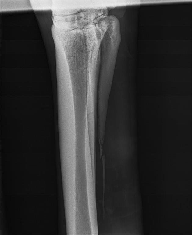
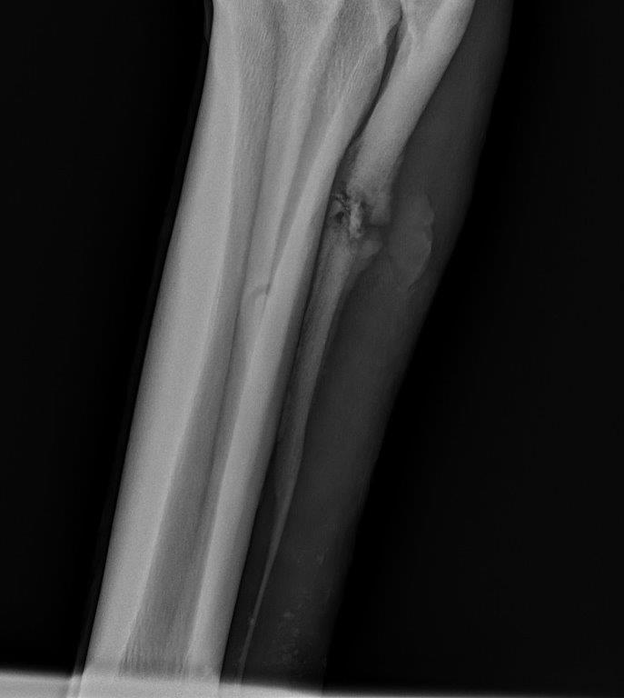
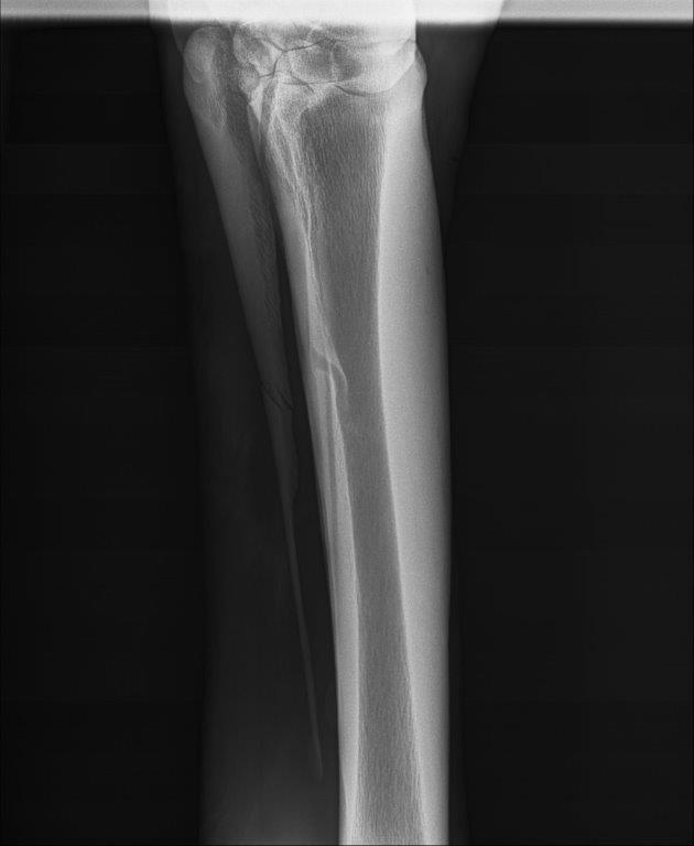
Fractures to the splint bones are not uncommon and can occur as the result of direct trauma to the bone or can occur when excessive swelling occurs within the limb, pushing the lower part of the splint outwards, causing it to snap with the pressure. In some cases, tension in the interosseous ligament will cause the distal part of the splint to bow and fracture during exercise when the fetlock drops (hyperflexion of the fetlock).
In my experience, fractured lateral or outside splint bones generally occur through trauma, frequently when kicked by another horse, or where they kick a fixed or solid object. Fractured medial or inside splint bones can occur with trauma but can also be attributed to swelling or tension in the soft tissues as described above, that forces the splint to curve outwards from the body and snap the bottom of the bone off.
“Fractures to the splint bones
are not uncommon.”
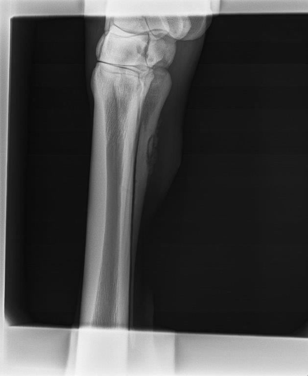
Diagnosis of a fractured splint is usually a simple process involving an X-ray of the affected site, but in cases where the suspensory ligament is swollen, fractures can be visualised on the ultrasound examination if the veterinarian images the bone. Sometimes when a wound is present, any swelling or pain is attributed to the wound and the potential for the splint bone to be involved is overlooked. If the wound has healed and the swelling remains, or in some cases the wound does not heal and there is a persistent discharge, this should alert the vet to the underlying issue and the need to X-ray the limb. Occasionally, I have been able to diagnose a fractured splint bone when examining a wound, as I am able to feel the pieces of fractured bone within the wound, however, it is always better to X-ray the splint bone to assess the type and severity of the fracture as this can change the treatment plan.

Depending on the location of the fracture, and whether an open wound is present or not, treatment of splint bone fractures can vary. For fractures in the lower part of the splint bone, either medially or laterally, the fragment can be removed easily and prognosis for returning to competition is good. If left to heal alone, many of these will become sound but the cosmetic result may not be as good. The important thing to assess with these distal fractures is the cause of the fracture. If the splint bone has fractured because of a suspensory ligament injury, the treatment plan including time out and prognosis on return to competition is governed by the damaged suspensory ligament and not the fracture.
For fractures that occur in the proximal portion (upper third), removing the splint fragment is less desirable. Fractures that are not associated with an open wound generally heal with conservative treatment alone, although occasionally a sequestrum may form and surgery is required to remove the dead piece of bone. For fractures with an open wound, the risk of the splint bone becoming infected is high and surgery may be required to remove fragments of bone and curette out areas of infection to allow the wound to heal. Some cases of open wound fractures can be treated conservatively, using daily lavages and antibiotics to clean up the infection, but this can take a prolonged period.
For some proximal fractures where removal of the bone is not desirable, internal fixation using a plate and screws can be performed. A plate and screws can be used to stabilise the fracture to allow the splint to heal, or they can be used to secure the proximal portion of the splint bone to the cannon bone, should it be necessary to remove a large part of the splint. Implants, however, when placed into infected sites will frequently become a source of ongoing infection and need to be removed within weeks of being placed. Therefore, some infected wounds that involve the proximal aspect of the splint are left to heal without fixation of the fragment to avoid having to remove the hardware early. For some of the fractures in the middle section of the splint bone, a segment of bone is removed, leaving the proximal portion and the lower portion of the splint in situ.
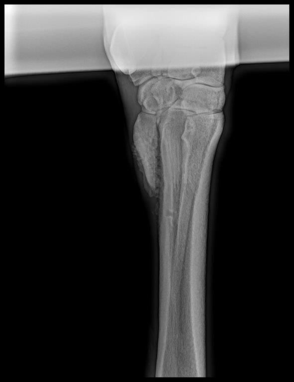
The horse produces a bony callous to help repair many types of fractures in the body, but sometimes these callouses can become excessively large, especially if there is movement between fragments or the fragments have become displaced, leaving a space between the fragments that the body tries to bridge. This can be particularly true of splint bone fractures, causing large unsightly lumps if the callous develops on the outside of the limb, or lameness and suspensory ligament problems if they develop on the inside of the splint bone. When the callous becomes large, it can rub on the suspensory ligament, damaging the fibres and causing a chronic lameness.
This can occur when the fragments are apart and left to heal conservatively (if the callous is formed trying to connect the distal part of the fracture to the proximal section), or on the end of the remaining piece of splint bone when the lower part of the splint bone has been removed but instability exists. Further surgery is then required to remove the callous. In these cases, it is very important to reduce the risk of further excessive callous developing by removing some of the inciting factors, such as the removal of the distal fragment and/or stabilising the remaining splint bone. The horse will also need to be well bandaged, and box rested to minimise movement.
There are other complications associated with the removal of a large amount of the splint bone and one of these is subluxation of the splint bone. A subluxated splint bone has no solid attachment to the cannon bone and therefore loses the support it gives to either the carpus or hock bone that sits on the base of the corresponding splint bone. This occurred recently with me and resulted in a very lame horse. A three-year-old filly had sustained a kick to her right front lateral splint and had surgery to remove most of the splint bone due to an open wound that extended into the fractured area of bone (I could readily palpate pieces of fractured splint at the time of presentation).
Surgery all went to plan but a week after returning home, the filly started to become progressively lame to the point she was walking on three legs when asked to move. With the worry of an infection being the major concern, multiple tests including blood tests and a joint tap of the knee were performed to look for any sources of infection. As all results came back negative for infection, and subluxation of the splint bone and fourth carpal bone was the likely diagnosis, causing severe pain when the filly walked or turned. Treatment involved tight bandaging and bracing of the knee and upper limb to minimise movement between the bones. She was unable to have the remaining splint fixed to the cannon as the wound was opened and no implant could safely be used. Over several weeks, the body formed a large callous around the area and this stabilised the splint by fusing it to the cannon bone. Currently the filly is walking a lot better, however, it is too early to say whether this callous will continue to grow and cause further complications down the track. EQ
YOU MIGHT ALSO LIKE TO READ BY DR MAXINE BRAIN:
When Horses Choke – Equestrian Life, June 2023
The Challenge of Treating HPSD – Equestrian Life, May 2023
From the Horse’s Mouth: Salivary Glands – Equestrian Life, February 2023
Cardiac Murmurs – Equestrian Life, February 2023
Matters of the Heart – Equestrian Life, January 2023
Umbilical Concerns in Foals – Equestrian Life, December 2022
Retained Foetal Membranes – Equestrian Life, October 2022
Preparing for Laminitis – Equestrian Life, September 2022
Working Together for Best Outcomes – Equestrian Life, August 2022
What Constitutes an Emergency – Equestrian Life, July 2022
Peri-Tarsal Cellulitis Calls for Quick Action – Equestrian Life, June 2022
Sinusitis: Not To Be Sneezed At – Equestrian Life, May 2022
Japanese Encephalitis: No Cause For Alarm – Equestrian Life, April 2022
Hernia Learning Curve – Equestrian Life, March 2022
Osteochondromas: Benign But Irritating – Equestrian Life, February 2022
Don’t Forget the Water – Equestrian Life, January 2022
Understanding Anaesthesia – Equestrian Life, December 2021
A Quick Guide to Castration – Equestrian Life, November 2021
Caring for Mammary Glands – Equestrian Life, October 2021
Sepsis In Foals – Equestrian Life, September 2021
Understanding Tendon Sheath Inflammation – Equestrian Life, August 2021
The Mystery of Equine Shivers – Equestrian Life, July 2021
Heads up for the Big Chill – Equestrian Life, June 2021
The Ridden Horse Pain Ethogram – Equestrian Life, May 2021
The Benefits of Genetic Testing – Equestrian Life, April 2021
Heavy Metal Toxicities – Equestrian Life, March 2021
Euthanasia, the Toughest Decision – Equestrian Life, February 2021
How to Beat Heat Stress – Equestrian Life, January 2021
Medicinal Cannabis for Horses – Equestrian Life, December 2020
Foal Diarrhoea Part 2: Infectious Diarrhoea – Equestrian Life, November 2020
Foal Diarrhoea (Don’t Panic!) – Equestrian Life, October 2020
Urticaria Calls For Detective Work – Equestrian Life, September 2020
Winter’s Scourge, The Foot Abscess – Equestrian Life, August 2020
Core Strengthening & Balance Exercises – Equestrian Life, July 2020
The Principles of Rehabilitation – Equestrian Life, June 2020
When is Old, Too Old? – Equestrian Life, May 2020