Horses are commonly affected by respiratory complaints, with asthma, nasal discharges, coughing, bleeding, and pneumonia among the main ones we see.
Horses involved in athletic disciplines are more likely to be referred to a veterinarian for a diagnosis as symptoms are generally more obvious when exercising and can interfere with the plans and expectations owners have for the horse. Whilst not all respiratory problems require veterinary intervention, the practice of calling the vet to treat coughs and respiratory issues routinely with antibiotics is now being frowned upon due to the emergence of antibiotic resistance. Instead, the emphasis is moving towards diagnosing the respiratory disorder correctly, therefore allowing the problem to be treated appropriately.
There are many tools a veterinarian can use to investigate the cause of a respiratory complaint. The first is one that has been around for decades but is often underrated as a diagnostic tool, and this is the stethoscope. Performing a clinical examination is very important when assessing a horse for any ailment and the stethoscope is essential for performing a thorough physical examination. For respiratory examinations, the stethoscope is used for listening to the chest to identify any wheezes or crackles, locate areas of affected lung and alert the vet to the presence of any fluid that may be present. The vet can also check the heart rate and rhythm whilst examining the lungs to eliminate the heart as a cause of a respiratory complaint and to ascertain if the respiratory ailment is systemically affecting the horse.
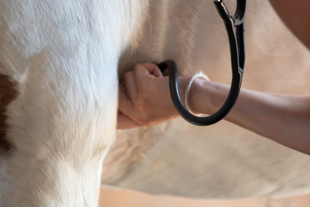
“The stethoscope is
essential for performing
a thorough physical examination.”
Some vets will use a rebreathing bag whilst listening to the chest with a stethoscope. This is basically a large bag held over and above the nose and mouth that restricts the amount of fresh air the horse can breathe in. By rebreathing the same air, the carbon dioxide (CO2) increases, and oxygen decreases within the bag, reducing the oxygen available within the lungs and increasing the CO2 in the bloodstream. This triggers the receptors that stimulate the horse to inhale larger volumes of air. When the horse inhales large volumes of air, it is much easier to auscultate abnormal lung sounds. (Hence why in humans the doctor says to take a deep breath when they listen to the chest.)
“Most horses tolerate
this procedure well…”
The next procedure most veterinarians will undertake when asked to assess the respiratory system is endoscopy, commonly referred to as a “scope”. This is a very common procedure and involves passing the scope (a thin tube with a camera on the end) up through the nasal passage, into the pharynx (throat) and down into the trachea to the bifurcation of the lungs. Whilst in the pharynx, various structures in the throat, such as the larynx and soft palate, are assessed for any abnormalities, either structural or functional. The scope is passed through the larynx into the trachea to look for any blood or mucus that may be negatively affecting the horse. Although horses will generally cough when the scope moves into the trachea, most horses tolerate this procedure well.
In cases where a functional abnormality is suspected but not visualised when the horse is scoped at rest, the veterinarian may need to use a dynamic scope. The standard endoscope only shows how the larynx appears at rest and how it functions when breathing and swallowing. For horses that have respiratory abnormalities that generate respiratory noises or poor performance associated with exercise, a scope is placed into the nose and positioned so that the pharynx and larynx can be viewed while the horse is working. Some machines allow the image to be viewed as the horse works, whilst others rely on the images being recorded onto an SD card and watched on a computer afterwards to diagnose functional abnormalities.
After the physical and/or scope examination has been performed, the vet may suspect there is fluid on the horse’s chest and decide to do an ultrasound examination of both sides of the chest. The ultrasound is a great tool for examining any abnormality on the outside surface of the lung lobe and would be the tool of choice to examine horses with pleuropneumonia. The ultrasound examination shows if there has been fluid accumulation between the chest wall and the lung as well as identifying any areas of lung consolidation. It is important to assess both sides of the chest as the two sides don’t always communicate and it is possible to have fluid on one side of the chest and not on the other.
Unfortunately, there are limitations with ultrasound imaging of the lung as ultrasound waves will not pass through air, and therefore only the surface of normal lung can be visualised. This means that abscesses or tumours deep in the lungs will not be detected unless they are associated with changes occurring on the lung surface.
X-rays are of limited value when it comes to the respiratory system. A horse’s chest is far too large for standard ambulatory X-ray machines to penetrate. The exceptions to this are foals and miniature horses where X-rays may be used to look for abscesses or other densities in the lung. X-rays can be used for the upper respiratory tract to assess the nasal passages, sinuses, pharynx, larynx, and trachea and are required to identify masses and fluid in areas that cannot be adequately examined using the endoscope.
CT scans are quickly superseding X-rays of the head due to the superior images and detail that can be acquired, which then allows for a better diagnosis. Currently, however, CT machines are restricted in their availability and are usually only found in universities and large equine specialist practices. In time, CT units may become increasingly available around the country, encouraging their use on a more routine basis. Practices with access to MRI machines are also using these to image the upper airways rather than using X-rays because they provide a much greater diagnostic image. As an MRI of the head often requires general anaesthesia to perform, its use is restricted to more involved cases.
Scintigraphy can be used in some specialty equine veterinary hospitals to assess how areas of the lung are perfused with blood and how well-ventilated different lobes of the lung are. Using nuclear medicine for respiratory ailments, however, would be an uncommon procedure to perform in Australia.
LABORATORY TESTS
Laboratory assessments for investigating respiratory problems are very common and are often used as part of a routine diagnostic workup. Blood tests can be useful in cases of infection, allowing the vet to monitor the course of an infection by tracking inflammatory markers like fibrinogen and serum amyloid A (SAA). They are, however, of limited value for many respiratory abnormalities as they give a picture of the overall health of the horse but are not useful in diagnosing common conditions such as Inflammatory Airway Diseases or Equine Asthma.
One of the more common diagnostic tests performed on the lungs is a bronchoalveolar lavage (BAL), colloquially known as a “lung wash”. This is a non-invasive procedure that can be done at the stables or on the farm and allows the veterinarian to collect cells from the lower airways and look for evidence of infection, inflammation, or blood. The horse is sedated and a BAL tube is passed through the nose, down the trachea and into a segment of lung where it is wedged gently into a bronchiole. A quantity of sterile fluid is inserted through the tube and into the segment of lung and then aspirated back up through tube and collected. The idea is the sterile fluid washes a small area of the lung, flushing numerous cells and mucus into the fluid, which is then sent to a laboratory for analysis. The cell types are then quantified and assessed so a more informed diagnosis and treatment plan can be made.
This method is ideal for conditions that affect large areas of the lung such as asthma and chronic obstructive airway disease (heaves). It is also commonly used to assess horses that are suspected of having exercise induced pulmonary haemorrhage (EIPH) or “bleeding”. In these horses, a BAL is used to look for evidence of bleeding, as well as look for any underlying factors that may have contributed to the bleed. Some horses will have an underlying infection or allergy that has weakened or inflamed the lung tissue, increasing the likelihood and severity of the horse bleeding when exercised. By identifying these underlying factors, better treatment protocols can be put in place to reduce the risk of further haemorrhage occurring.
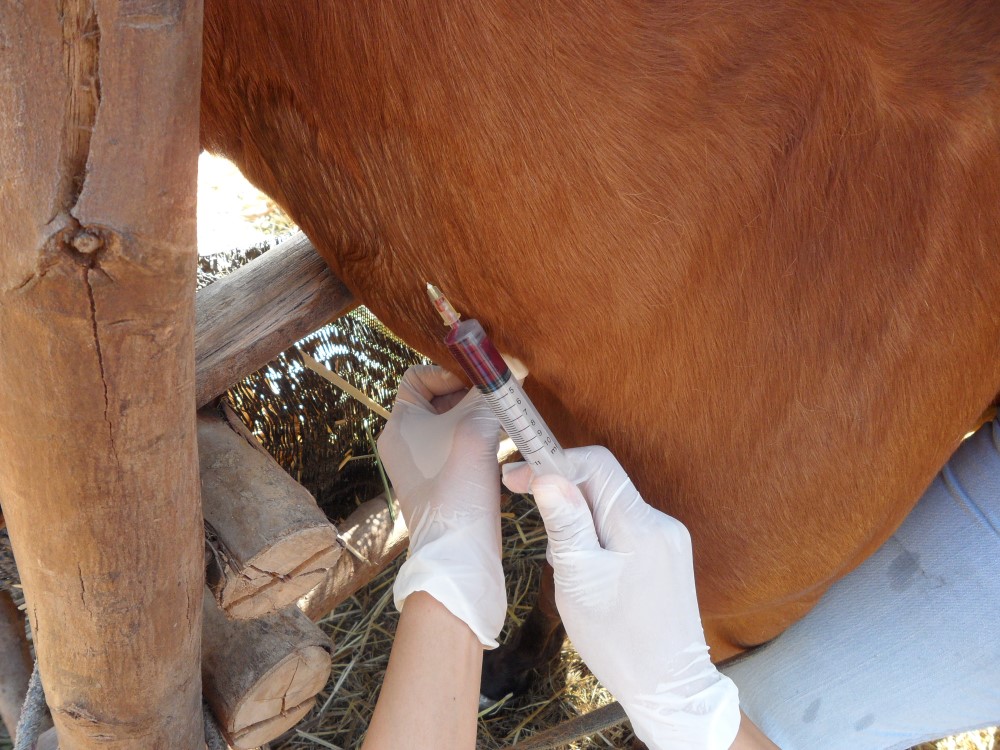
A transtracheal wash (TTW) is another common diagnostic procedure that can be used to further investigate lung infections or mucus. This is ideal when looking to isolate the bacteria involved in a pneumonia so appropriate antibiotics can be used.
There are two ways of performing a TTW, one is non-invasive and is done via a small tube passed down through a port in the endoscope. Once in the trachea, the tube is pushed out past the end of the scope and a small quantity of sterile fluid flushed through the tube into the trachea. This fluid runs a small distance along the trachea, washing down mucus and cells into a puddle towards the base of the trachea. From here, it is aspirated back up through the tube and collected in a sterile pot that is then sent to the lab. The fluid is assessed with a microscope, and then cultured to identify any causative bacteria before sensitivity testing is performed to select a suitable antibiotic to treat the offending bacteria.
The downside of this procedure is there is a risk of contamination of the sample occurring and this will obscure the true test results. As the scope passes through the nose and upper airways (which are not sterile environments), resident bacteria in the pharynx can become adhered to the tip of the scope and be incorporated into the fluid as the sample is collected.
The second way to perform a TTW is more invasive and involves inserting a large-bore, sterile needle into the trachea through a surgically prepared site (clipped and scrubbed) on the lower aspect of the neck. A thin sterile tube is then passed through the needle into the trachea and fluid inserted and retrieved in a similar manner to the aforementioned TTW. Whilst this is a better way to collect a tracheal sample as it bypasses the nasal passages and pharynx, there is a higher risk of complications occurring compared to collecting a sample via the endoscope.
Using the second method can cause cellulitis and abscess formation at the site of needle placement into the trachea. This is due to the tube becoming contaminated with bacteria in the trachea, so when the tube is pulled back through the skin, these bacteria seed into the soft tissues of the neck, and cause an infection.
Polymerase chain reaction testing, better known as PCR tests, are laboratory tests used to confirm the presence of viruses, bacteria, rickettsia, and fungi by identifying the pathogens in a DNA sample taken from the patient. For horses, there are several respiratory pathogens that can be identified or eliminated as causes of infection using PCR on a swab of the nasopharynx. A large swab is passed up through the nose, into the pharynx and swirled about to collect a sample that is placed into a special media and sent for testing. Depending on the lab, only certain viruses or bacteria can be screened for and these can include Equine Influenza virus, Equine Herpesvirus, Equine Rhinitis, Equine Adenovirus and Streptococcus Equi sub sp equi (strangles).
Thoracocentesis is the placement of a needle or catheter into the chest to obtain a sample of fluid from the space between the lung and the chest wall, after it has been diagnosed on ultrasound. The sample is assessed for cell types (cytology) and cultured to isolate the causative bacteria in cases of pleuropneumonia. Generally, after a sample is taken for diagnostic purposes, the fluid is drained out of the chest via the same portal as part of the treatment.
A lung biopsy can be used when abnormal lung tissue or an abnormal mass is identified on an ultrasound examination. It is not a common procedure but can be performed on site using the ultrasound to guide the biopsy instrument to the appropriate site.
Whilst some research hospitals and universities have access to pulmonary function tests to evaluate how effectively the lungs are working, these tests are not readily available to horse owners in Australia. These tests can be performed on horses at rest and in some circumstances whilst exercising on a treadmill. Parameters assessed include air flow in and out of the lungs, respiratory system resistance, pleural pressures, and gas analysis on the blood. Assessing lung function and performance limitations in horses, however, is perhaps one area of respiratory diagnosis that we need to improve in, as many of the available procedures are invasive and therefore not acceptable to many horse owners.

A healthy respiratory system for the horse is vital and maintaining our horses in tip top order relies on our abilities to identify and manage respiratory irregularities. There are many procedures available to help diagnose respiratory disorders in our horses, so it is well worth discussing different options with your veterinarian before deriving any treatment plans that aren’t based on a sound diagnosis. EQ
YOU MIGHT ALSO LIKE TO READ BY DR MAXINE BRAIN:
Anhidrosis: What Is Is? – Equestrian Life, January/February 2024
Fractured Jaws – Equestrian Life, December 2023
Keeping Our Country Free of Disease – Equestrian Life, November 2023
Managing Endometritis – Equestrian Life, October 2023
Granulosa Cell Tumours – Equestrian Life, September 2023
Being a Horse in Africa – Equestrian Life, August 2023
Splint Bone Fractures – Equestrian Life, July 2023
When Horses Choke – Equestrian Life, June 2023
The Challenge of Treating HPSD – Equestrian Life, May 2023
From the Horse’s Mouth: Salivary Glands – Equestrian Life, February 2023
Cardiac Murmurs – Equestrian Life, February 2023
Matters of the Heart – Equestrian Life, January 2023
Umbilical Concerns in Foals – Equestrian Life, December 2022
Retained Foetal Membranes – Equestrian Life, October 2022
Preparing for Laminitis – Equestrian Life, September 2022
Working Together for Best Outcomes – Equestrian Life, August 2022
What Constitutes an Emergency – Equestrian Life, July 2022
Peri-Tarsal Cellulitis Calls for Quick Action – Equestrian Life, June 2022
Sinusitis: Not To Be Sneezed At – Equestrian Life, May 2022
Japanese Encephalitis: No Cause For Alarm – Equestrian Life, April 2022
Hernia Learning Curve – Equestrian Life, March 2022
Osteochondromas: Benign But Irritating – Equestrian Life, February 2022
Don’t Forget the Water – Equestrian Life, January 2022
Don’t Forget the Water – Equestrian Life, January 2022
Understanding Anaesthesia – Equestrian Life, December 2021
A Quick Guide to Castration – Equestrian Life, November 2021
Caring for Mammary Glands – Equestrian Life, October 2021
Sepsis In Foals – Equestrian Life, September 2021
Understanding Tendon Sheath Inflammation – Equestrian Life, August 2021
The Mystery of Equine Shivers – Equestrian Life, July 2021
Heads up for the Big Chill – Equestrian Life, June 2021
The Ridden Horse Pain Ethogram – Equestrian Life, May 2021
The Benefits of Genetic Testing – Equestrian Life, April 2021
Heavy Metal Toxicities – Equestrian Life, March 2021
Euthanasia, the Toughest Decision – Equestrian Life, February 2021
How to Beat Heat Stress – Equestrian Life, January 2021
Medicinal Cannabis for Horses – Equestrian Life, December 2020
Foal Diarrhoea Part 2: Infectious Diarrhoea – Equestrian Life, November 2020
Foal Diarrhoea (Don’t Panic!) – Equestrian Life, October 2020
Urticaria Calls For Detective Work – Equestrian Life, September 2020
Winter’s Scourge, The Foot Abscess – Equestrian Life, August 2020
Core Strengthening & Balance Exercises – Equestrian Life, July 2020
The Principles of Rehabilitation – Equestrian Life, June 2020
When is Old, Too Old? – Equestrian Life, May 2020